OsteoBiol by Tecnoss
INTERNATIONAL HANDS-ON
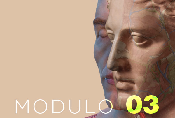
HUMAN CADAVER COURSE


OsteoBiol by Tecnoss

The course deals with the application of the main basic and advanced techniques of regenerative surgery around dental implants, with associated dissection on cadaver of the related muscle and neurovascular tissues.
Starting from the elevation of full-thickness and partial-thickness flaps, the described surgical techniques will be performed first, followed by dissection of the anatomical structures surrounding the surgical area.
The dissection taught will be a “clinically oriented” dissection by planes, that is to say structured with the objective of understanding, through the acquisition of the anatomical data, how to prevent and manage haemorrhage and neurosensorial complications, intra- and postoperatively respectively.
This approach is essential to dispel doubts and fears during the application of the techniques, while increasing your clinical skills.
The course is conducted on anatomical preparations of fresh-frozen cadavers that ensure maximum safety for the participants from the point of view of infections and provide an ideal superimposability to the living person.
85% of the course will be focused on practice, the keyword will be “do” and the more theoretical aspects will be combined with hands-on demonstrations.
One head for every 2 participants will be made available to give everybody the opportunity to take part directly.

2 giorni di formazione
5-6 aprile 2024
Registration fee includes two alternative bus transfers from BGY airport on 26th afternoon, all lectures and hands-on and Friday social dinner.
Registrations fee € 3.000 (22% VAT included)
Social dinner on Friday evening at Ristorante “Il Violino” in Cremona: www.ilviolino.it
CANCELLATIONS
50% reimbursement if cancellation is made before November 30th, 2022
No reimbursement after November 30th, 2022
In case of course cancellation due to new Covid restrictions or for not having reached the minimum number of participants, all pre-paid fees will be completely reimbursed.
No flight tickets or hotel bookings will be reimbursed in case of course cancellation.
SECRETARIAT
Tecnoss Dental s.r.l.
Via Livorno 60 | 10144 Torino | Italy
Palazzo Trecchi | Via Sigismondo Trecchi 20 | 26100 Cremona | ITALY
RECOMMENDED HOTELS
Dellearti Design Hotel ★★★★ www.dellearti.com
Hotel Continental ★★★★ www.hotelcontinentalcremona.it
TRAVEL INFORMATION
Private minibus transfers from BGY airport to Cremona on Thursday 26th January 2023 at 16:00 and 19:00 hours only: transfer time approximately one hour; no other group transfers will be organized nor included in the course fee at different times or from other airports.
| 08:30 | Registration of participants |
|---|---|
| 09:00 | Welcome and introduction to the course |
| 09:15 | Theoretical teaching part with explanatory dissection and clinical videos: • anatomy of the mandible and of muscular and neurovascular structures in relation to the treated surgical techniques • overview of the surgical techniques to be performed |
| 10:30 | Hands-on cadaver with application of surgical techniques in the mandibular area and dissection of the relative muscular and neurovascular structures of major interest (1° stage):
Anatomical Dissection |
| 13:30 | Lunch: catering service at Trecchi Palace |
| 14:30 | Hands-on cadaver with application of surgical techniques in the mandibular area and dissection of the relative muscular and neurovascular structures of major interest (2° stage):
Surgical Techniques |
| 08:30 | Registration of participants |
|---|---|
| 09:00 | Theoretical teaching part with explanatory dissection and clinical videos: • anatomy of the maxilla and of muscular and neurovascular structures in relation to the treated surgical techniques • overview of the surgical techniques to be performed |
| 10:30 | Hands-on cadaver with application of surgical techniques in the maxillary area and dissection of the relative muscular and neurovascular structures of major interest (1° stage):
Anatomical Dissection |
| 13:30 | Lunch: catering service at Trecchi Palace |
| 14:30 | Hands-on cadaver with application of surgical techniques in the maxillary area and dissection of the relative muscular and neurovascular structures of major interest (2° stage):
Surgical Techniques |
| 08:30 | Registration of participants |
| 09:00 | Theoretical teaching part with explanatory dissection and clinical videos: • anatomy of the maxilla and of muscular and neurovascular structures in relation to the treated surgical techniques • overview of the surgical techniques to be performed |
| 10:30 | Hands-on cadaver with application of surgical techniques in the maxillary area and dissection of the relative muscular and neurovascular structures of major interest (1° stage):
Anatomical Dissection |
| 13:30 | Lunch: catering service at Trecchi Palace |
| 14:30 | Hands-on cadaver with application of surgical techniques in the maxillary area and dissection of the relative muscular and neurovascular structures of major interest (2° stage):
Surgical Techniques |
Studia l’anatomia del distretto oro-maxillo-facciale sia a livello intra-orale che extra-orale e approfondisci la conoscenza di quelle variabili anatomiche maggiormente associate a complicanze intra- e post-operatorie discusse nell’ambito della letteratura scientifica internazionale attraverso una didattica scientificamente orientata verso l’evidence based.
Iscrivendoti ai corsi dell’Academy of Craniofacial Anatomy potrai:
Che cos’è l’Academy of Craniofacial Anatomy?
L’Academy of Craniofacial Anatomy nasce nel maggio 2021 e come associazione culturale si propone lo scopo di promuovere lo studio e la ricerca scientifica dell’anatomia del massiccio facciale attraverso
Chi siete?
L’accademia si avvale di un corpo docente internazionale di anatomisti e clinici con ampia esperienza nell’ambito della dissezione anatomica e vanta di una solida partnership con docenti del dipartimento di anatomia cranio-cervico-facciale dell’Université René Descartes Paris 5, centro di riferimento storico per la dissezione anatomica a livello europeo e tra i più prestigiosi del mondo.
Ci onora dell’incarico di presidente onorario il Prof. Jean-François Gaudy, Professore Ordinario di Anatomia presso la Facoltà di Medicina e Chirurgia dell’Université René Descartes Paris 5, Parigi.
La vostra Mission?
L’obiettivo dell’Academy è quello di proporre una formazione UNICA NEL SUO GENERE con un approccio che deve essere prima da anatomista e poi da clinico; per questo verranno proposti dei corsi di anatomia dissettiva prima e poi di applicazione delle tecniche chirurgiche, proponendo moduli diversificati in base al livello e alle esigenze cliniche di ognuno.
Dove posso seguire i vostri corsi?
I corsi avranno luogo in una cornice decisamente esclusiva, ovverosia presso il Cadaver Lab di Palazzo Trecchi, edificio storico dal valore artistico e culturale inestimabile situato nel centro storico della città di Cremona e la cui fondazione risale al 1496.
Qual è il motto della vostra accademia?
Il motto della nostra accademia è “noi vediamo SOLO quello che conosciamo” e vuole proprio significare che se non si conosce una struttura anatomica nel dettaglio sarà di certo improbabile prevenire e gestire le complicanze ad essa correlate.
Grazie alla dissezione anatomica si toccano con mano le strutture muscolari e neuro-vascolari di interesse e le loro variabilità anatomica, aspetto questo fondamentale per la crescita del chirurgo nell’ottica di imparare a gestire e soprattutto prevenire complicanze emorragiche e neurosensoriali.
Cosa si intende per dissezione anatomica?
Da non confondere i termini “dissezione anatomica” e “autopsia” che non sono affatto sinonimi: mentre l’autopsia è una pratica eseguita dal medico legale al fine di individuare la/le cause di morte, e dunque non necessariamente conservativa, la dissezione anatomica è una pratica altamente conservativa eseguita da un Anatomista, basata sulla separazione dei diversi piani e la visualizzazione dei rapporti tridimensionali tra le singole strutture.
Nonostante le innovative metodiche d’indagine radiologica e la continua evoluzione delle tecniche chirurgiche, rimane centrale lo studio dell’anatomia dissettiva; sono gli stessi operatori in campo chirurgico a ritenere che l’esperienza diretta sul cadavere sia insostituibile e che la dissezione anatomica rivesta un’importanza fondamentale nella formazione di specializzandi e nell’aggiornamento degli specialisti.
Perché la dissezione anatomica è così importante?
La dissezione anatomica è lo strumento principe di insegnamento e apprendimento per conoscere il corpo umano attraverso l’esperienza diretta, differenziandosi dall’autopsia e dalla pratica chirurgica.
E’ stata anche definita da Dyer & Thorndike (2000) come “il passaggio universalmente riconosciuto per diventare medico”.
Infatti, solo attraverso la formazione su cadavere l’anatomia da materia di studio mnemonico sui libri diventa un vero e proprio specchio della realtà applicativa, consentendo al clinico di acquisire il senso della consistenza dei tessuti, di individuare direttamente i particolari anatomici più complessi e di apprendere i rapporti tridimensionali di vasi, nervi e organi nelle diverse regioni del corpo.
È previsto un preparato anatomico per ciascun discente?
Sarà messa a disposizione una testa ogni 2 partecipanti per dare a tutti la possibilità di essere sempre operativi in prima persona.
Il partecipante effettuerà in prima persona la dissezione delle strutture anatomiche trattate e applicherà le tecniche chirurgiche descritte direttamente sul cadavere utilizzando lo strumentario e i materiali messi a disposizione.
In che percentuale nei corsi ACA si darà rilevanza alla parte pratica?
La parte pratica rappresenterà l’80% della corsistica, la parola chiave sarà “fare” e l’aspetto didattico più teorico sarà spesso contestuale alle dimostrazioni pratiche.
Che tipo di riconoscimento ottiene chi partecipa ai corsi?
Ogni corsista otterrà l’attestato di partecipazione al programma formativo dell’Academy e per ciascun modulo verranno rilasciati i relativi crediti ECM erogati dal Ministero della Salute.
Quanto costano i corsi?
I nostri corsi hanno costi variabili; puoi consultare la pagina di ciascun modulo per avere informazioni sui costi.
Posso rateizzare o finanziare la quota di partecipazione?
Assolutamente si! La nostra segreteria ti darà tutte le informazioni per accedere alle proposte di rateizzazione e finanziamento.